Moderate Hip dysplasia – HD “D”
Moderate Hip dysplasia – HD “D”
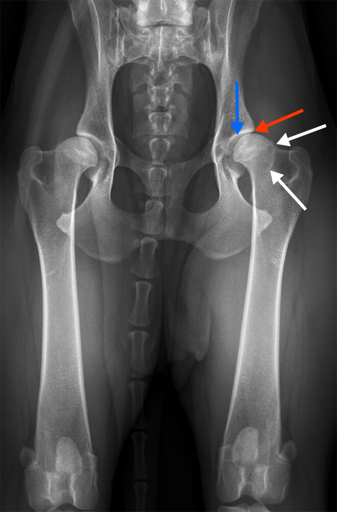
X-ray of the pelvis in the ventrodorsally optical path (position 1)
Typical example of moderate HD / HD “D”
Divergent joint space (blue arrow), slightly deformed femoral heads with flat acetabula, the cranial border of which has an externally significantly broadened subchondral bone plate has (red arrow) significant bony formations (osteophytes) on both femoral necks as a sign of existing arthrosis (white arrow). Wide “Morgan Line” (white arrow).
The Norberg angle is about 90° on both sides (normally 105∞).