FCP/ Coronoid Disease


FCP/ Coronoid Disease
The Ulna’s Coronoid Process disease - Processus Coronoideus Medialis Ulnae (PCM).
Forms: Fracture, fissure, atrophy of the bone
Fig. 1
Regular PCM (blue arrows) for comparison in lateral/ mediolateral view.
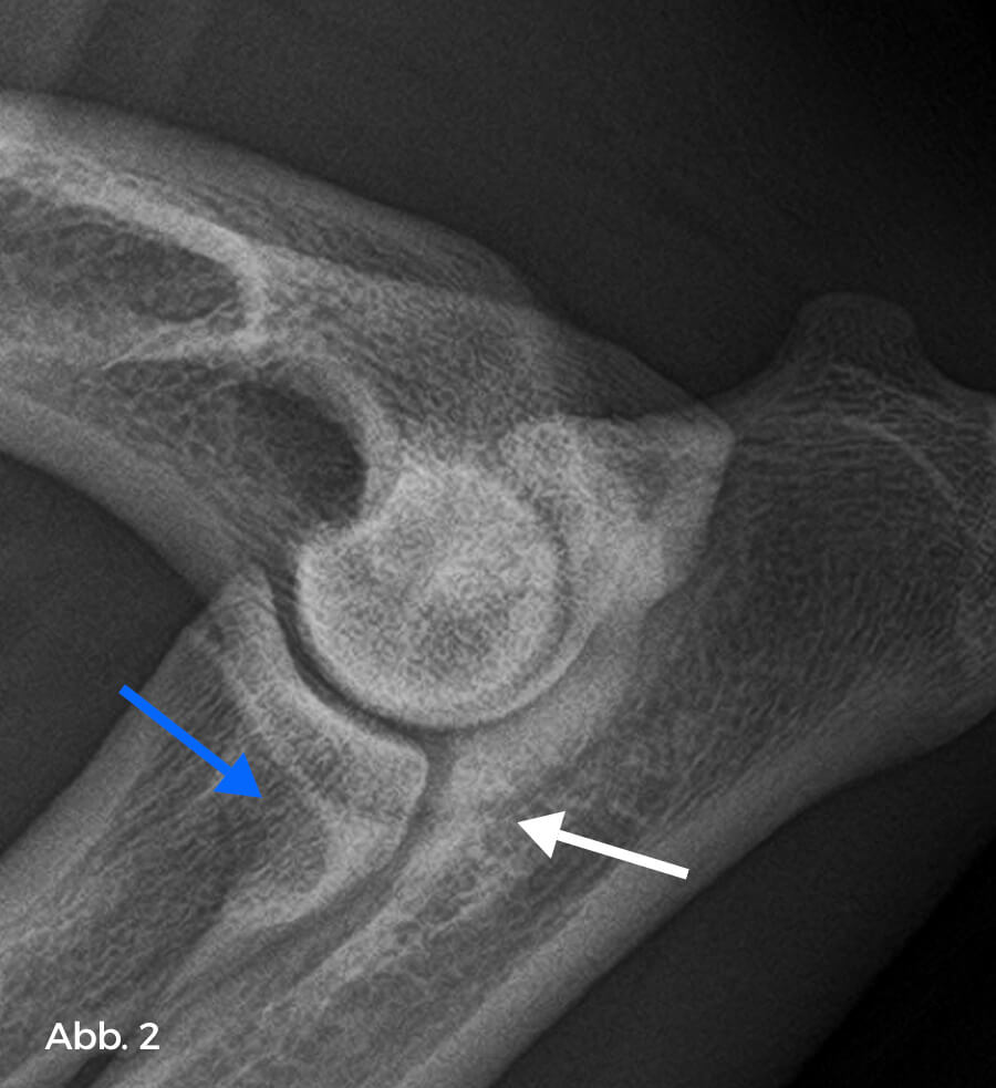
Fig. 2
Defect in the area of the tip of the PCM (blue arrow) with sclerosis at the base (white arrow). Although not always directly visible, a typical finding is the presence of a fragment. The direct evidence of a fragment is rarely visible (craniocaudal 15° pronated, fig. 3 and 4).
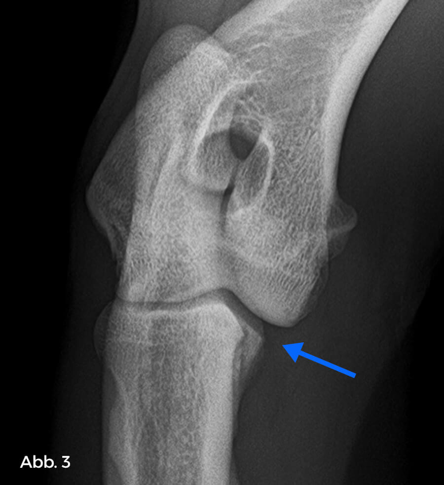
Fig. 3:
Example of no visible fragment (blue arrow)
Fig. 4:
Example of direct evidence of a fragment (blue arrow), including a subchondral defect in the trochlear joint surface (white arrow)